Capabilities
CyTOF® XT System allows researchers to dive deep into single-cell proteomics by detecting over 50 markers
Technology
CyTOF® XT System allows researchers to dive deep into single-cell proteomics by detecting over 50 markers
Workflow
CyTOF® XT System allows researchers to dive deep into single-cell proteomics by detecting over 50 markers
Overview
Unlocking New Insights in Tissue Microenvironments
Understanding the intricate behavior of individual cells is critical in today’s biomedical research, especially for immune system analysis, disease progression, and therapeutic response.
Imaging Mass Cytometry™ (IMC™)
The dynamics of tissue microenvironments play a crucial role in understanding disease progression, therapeutic response, and biomarker discovery. Imaging Mass Cytometry™ (IMC™) technology, powered by the Hyperion™ XTi Imaging System, enables researchers to explore the complexity of these dynamics with unparalleled precision. By detecting over 40 protein and RNA markers simultaneously in tissue samples, IMC uncovers spatial relationships that conventional imaging methods overlook.
Designed for high throughput and reproducibility, IMC offers the highest dynamic range in the industry, making it ideal for translational and clinical research. Whether focusing on oncology, immunology, or other complex diseases, these spatial proteomics solutions deliver the detailed insights needed to guide any phase of research.
Discover the throughput and precision that is uniquely designed for translational researchers.
Jana Fischer, PhD
Co-Founder and CEO, Navignostics
Capabilities
Unparalleled Multiplexing Capability
Imaging Mass Cytometry™ (IMC™) is the most trusted technology that enables researchers to accurately assess complex phenotypes and immune spatial interactions in the tissue microenvironment.
High Dynamic Range
Unlike traditional fluorescence-based imaging techniques, which often struggle with signal saturation and autofluorescence, IMC captures both high and low levels of protein expression in the same sample with exceptional clarity.
Assay development time
Because IMC technology uses an antibody reagent cocktail, it is simple to mix and match different antibodies without extensive assay validation.
Simultaneous Detection of 40+ Markers
IMC allows for the simultaneous imaging of over 40 markers in a single tissue section, facilitating the detailed mapping of tissue architecture, immune cell infiltration, and biomarker expression. This multiplexing capability provides richer, more comprehensive data with fewer processing cycles than traditional methods.
Flexibility of tissue acquisition
Flexibility of tissue acquisition parameters (Hyperion imaging modes) and Automated, High-Throughput Imaging: With the Hyperion XTi™ Slide Loader, researchers can image up to 40 slides in 24 hours, making it easier to process large-scale studies and accelerate results without compromising precision – run an entire study in an automated, walkaway run.
No Autofluorescence Interference
IMC utilizes metal-tagged antibodies instead of fluorophores, eliminating issues with autofluorescence and delivering cleaner, more reliable data, even from challenging tissue samples.
Unraveling the Complexity of the Immune System
Fluorescence-Based Imaging
Traditional
Traditional imaging methods, such as cyclic immunofluorescence, are limited by their low dynamic range, autofluorescence interference, and slow, multi-cycle processes. For example, brightfield imaging (DAB) gives at best one order of magnitude (OoM) of dynamic range, while fluorescence gives 2–3 OoM and IMC gives 5 OoM. These limitations often result in incomplete data, as they cannot capture both high- and low-expressing biomarkers in a single image, leading to missed insights.
Our Solution
Fluorescence-Based Imaging
Standard Bio
IMC overcomes these challenges by capturing the full dynamic range of spatial biomarkers, detecting both high- and low-expressing proteins simultaneously. This approach eliminates the need for multiple imaging cycles and delivers high-resolution, quantitative data without the drawbacks of autofluorescence. IMC provides up to 100 times higher throughput than cyclic fluorescence, making it the ideal solution for large-scale research studies.
Staining Redundancy
< 20 Markers
Spectral Overlap
Technology
Imaging Mass Cytometry Technology
Imaging Mass Cytometry™ (IMC™) is the most trusted technology that enables researchers to accurately assess complex phenotypes and immune spatial interactions in the tissue microenvironment.
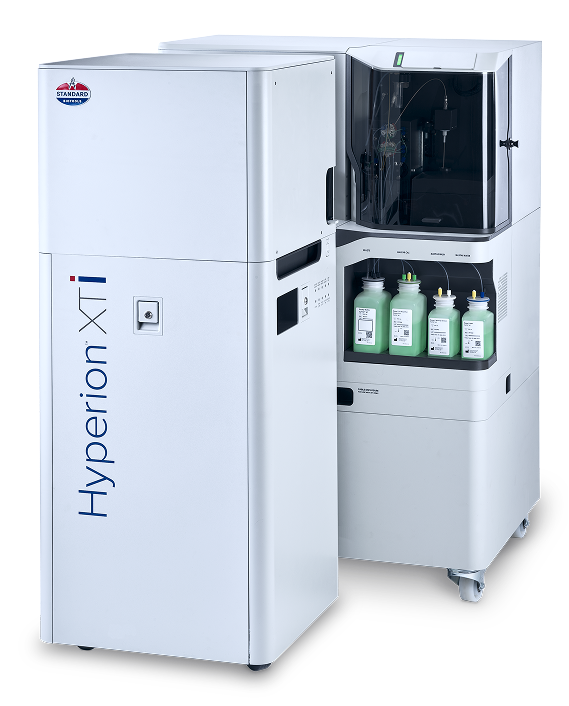
Hyperion XTi
CyTOF XT detects over 50 markers at once, far exceeding the limits of traditional flow cytometry. This enables a comprehensive analysis of immune populations and functional responses in a single experiment.
Discover the power of Imaging Mass Cytometry
Lorem ipsum dolor sit amet consectetur
Unique scale and high signal resolution
Simultaneously detect over 50 markers without spectral overlap
Higher-content, more reliable dataset in a single analysis
Faster, more accurate insights into complex cellular behaviors.
Our Solutions
Lorem ipsum dolor sit amet consectetur
Image any tissue type
with no autofluorescence interference
Image any tissue type
with no autofluorescence interference
Image any tissue type
with no autofluorescence interference
Image any tissue type
with no autofluorescence interference
Image any tissue type
with no autofluorescence interference
Image any tissue type
with no autofluorescence interference
Applications
Lorem ipsum dolor sit amet consectetur

Biomarker Discovery
Identify therapeutic targets that may be relevant to developing treatment strat…
Cell segmentation
See how the use of cell segmentation facilitates an end-to-end workflow for …
Dual Mode
Use your Hyperion XTi Imaging System to perform high-multiplex analysis of b…
Immuno-Oncology
Discover the impact of new immune therapies including checkpoint inhibit…
Dual Mode
Use your Hyperion XTi Imaging System to perform high-multiplex analysis of b…
Immuno-Oncology
Discover the impact of new immune therapies including checkpoint inhibit…
Case Studies
Lorem ipsum dolor sit amet consectetur
HER2 Trial, Clin. Cancer Res. 2019
Melanoma
IMC was used in a Phase 3 clinical trial of trastuzumab to stratify patients by HER2 expression levels, revealing how the dynamic range of HER2 expression and proximity to CD8+ T cells predicted patient response. Reference: Carvajal-Hausdorf, D.E. et al. 2019, Clinical Cancer Research.
HER2 Trial, Clin. Cancer Res. 2019
Breast Cancer
IMC was used in a Phase 3 clinical trial of trastuzumab to stratify patients by HER2 expression levels, revealing how the dynamic range of HER2 expression and proximity to CD8+ T cells predicted patient response. Reference: Carvajal-Hausdorf, D.E. et al. 2019, Clinical Cancer Research.
Checkpoint Therapy, Clin. Cancer Res. 2021
Melanoma
In a study of 60 patients with metastatic melanoma treated with immune checkpoint inhibitors, IMC identified 12 biomarkers of survival and response, including B2M, MHC-1, and LAG-3. Reference: Martinez-Morilla, S. et al. 2021, Clinical Cancer Research.
Hear how the team at Navignostics is translating spatial insights into the clinic by leveraging spatial proteomics into reporting within 72 hours of sample receipt, revolutionizing treatment decisions for each cancer patient.
Jana Fischer, PhD
Co-Founder and CEO, Navignostics
Explore
Resources and Data
Lorem ipsum dolor sit amet consec tetur adipisicing elit sed consec tetur adipisicing.
Overview
High-Throughput Automation
CyTOF XT automates much of the workflow, significantly reducing the hands-on time required by researchers and enabling the processing of large numbers of samples efficiently, without sacrificing data quality.
Overview
No Spectral Overlap:
Using metal isotopes rather than fluorescent dyes, CyTOF eliminates the common issue of spectral overlap found in traditional flow cytometry, providing cleaner and more precise data.
Overview
Comprehensive Immune Profiling
With the Maxpar® Direct Immune Profiling Assay™, researchers can measure 37 immune cell populations from a single tube of blood, providing detailed insights into immune function and facilitating faster, more informed decision-making in both preclinical and clinical settings.
Capabilities
Unparalleled Multiplexing Capability
CyTOF XT detects over 50 markers at once, far exceeding the limits of traditional flow cytometry. This enables a comprehensive analysis of immune populations and functional responses in a single experiment.
High Dynamic Range
Unlike traditional fluorescence-based imaging techniques, which often struggle with signal saturation and autofluorescence, IMC captures both high and low levels of protein expression in the same sample with exceptional clarity.
Assay development time
Because IMC technology uses an antibody reagent cocktail, it is simple to mix and match different antibodies without extensive assay validation.
Simultaneous Detection of 40+ Markers
IMC allows for the simultaneous imaging of over 40 markers in a single tissue section, facilitating the detailed mapping of tissue architecture, immune cell infiltration, and biomarker expression. This multiplexing capability provides richer, more comprehensive data with fewer processing cycles than traditional methods.
Flexibility of tissue acquisition
Flexibility of tissue acquisition parameters (Hyperion imaging modes) and Automated, High-Throughput Imaging: With the Hyperion XTi™ Slide Loader, researchers can image up to 40 slides in 24 hours, making it easier to process large-scale studies and accelerate results without compromising precision – run an entire study in an automated, walkaway run.
No Autofluorescence Interference
IMC utilizes metal-tagged antibodies instead of fluorophores, eliminating issues with autofluorescence and delivering cleaner, more reliable data, even from challenging tissue samples.
Discover the throughput and precision that is uniquely designed for translational researchers.
Jana Fischer, PhD
Co-Founder and CEO, Navignostics
Co-Founder and CEO, Navignostics